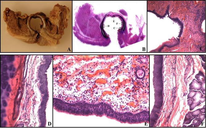
Figure 4.
Histopathology. A,B: The typical gross appearance of a cross-section at the stoma. A: The cross-section with fixation. B: A whole-mount large histological section showing the trachea, stoma, cutaneous mucosal junction, and the lateral muscle-trachea approximation. The boxes highlight areas illustrated at higher magnifications in the photomicrographs shown in C-F. C: The cutaneous-mucosal junction showing the transition from stratified squamous epithelium to respiratory mucosa. There is minimal chronic inflammation in both the subcutaneous tissue and the lamina propria of the trachea. Magnification 40X. D: The later tracheal wall. The respiratory mucosa has slight squamous metaplasia, but the lamina propria between the mucosa and cartilage has no pathological changes. Magnification 200X. E: The posterior tracheal wall opposite the stoma. Slight chronic inflammation is present in the lamina propria. This was the most severe chronic inflammation seen in any section of the trachea. The respiratory mucosa has no pathological changes. Magnification 200X. F: Lateral tracheal wall with squamous metaplasia, no chronic inflammation, and normal tracheal mucosal glands. There was neither an increase in tracheal mucosal gland thickness in the lamina propria, nor a pathological shift in the normal serous/mucous gland proportions. Magnification 200X.

