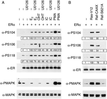
Figure 2.
Phosphorylation of S104 and S106 is stimulated by ERα ligands and by activators of MAPK. Immunoblots of lysates prepared from COS-1 cells transfected with empty expression vector (−) or expression vector for ERα, were performed as for Fig. 1. Lysates were additionally immunoblotted using antibodies for total (α-MAPK) and phosphorylated (α-PMAPK) Erk1/2 MAPK. Levels of phospho-ERα were quantitated in relation to the respective total ERα level (boxed, below each immunoblot). (A) Cells were pre-incubated with U0126 (10 μM) for 1 h, followed by the addition of E2 (10 nM), 4-hydroxytamoxifen (OHT, 100 nM), ICI 182 780 (ICI; 100 nM) or PMA (100 nM), as indicated, and cells harvested 30 min later. (B) Cells were transfected with expression vector for ERα, together with expression vectors for Ras-V12, Raf-CAAX or Raf-S621A, as indicated.

