
Figure 5. Loss of Rga4 Allows Bipolar Distribution of Active Cdc42 in Δpom1 Cells.
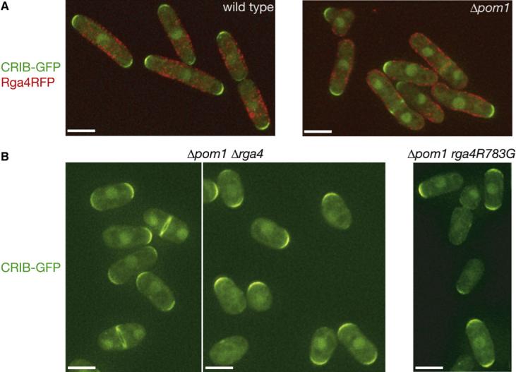
(A) Fluorescence microscopy of wild-type and Δpom1 strains expressing CRIB-GFP and Rga4RFP. (B) Fluorescence microscopy of Δpom1 Δrga4 and Δpom1 rga4R783G strains expressing CRIB-GFP. Immunoblotting confirmed that the Rga4R783G mutant protein was expressed at a level comparable to that of wild-type Rga4. Signals of CRIB-GFP and Rga4RFP were pseudo-colored green and red, respectively, and superimposed. Scale bars represent 5 μm. Images were taken at 0.4 μm steps and deconvolved for projection images.