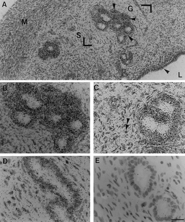
Figure 6.
In situ hybridization for uterus PAM mRNA in OVX or OVX+E2 rats. Cryostat uterus sections (10 μm) were hybridized to 35S-labeled antisense PAM riboprobe. Cryosections were dipped in photographic emulsion for histological resolution of autoradiographic grains. After 2 weeks of exposure, the tissues were processed and stained with hematoxylin. In situ hybridization signal in the uterus of the adult OVX rat (A) was observed in luminal (L) and glandular (G) cells (arrowheads); myometrium cells (M) were negative. The area indicated by the two right angles was magnified and shown in B. Stroma (S) cells appeared to be devoid of grains (see double arrowheads in C). (D) Compared with the OVX rats, labeling was generally less prominent in glandular cells of estrogen-treated OVX rats. (E) Bright-field micrograph of a section hybridized to sense PAM riboprobe shown for comparison. (The scale bar in E represents 25 μm for A and 50 μm for B–E.)

