
Fig. 8.
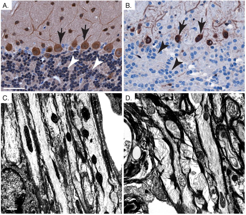
Pten loss in oligodendrocytes leads to cell autonomous defects in myelination in the cerebellum. A,B. Pten immunohistochemistry shows loss of Pten in granule neurons of the IGL (arrow head), and persistent Pten expression in Purkinje cells (arrows) from Pten cKO cerebellum (B) compared to control (A). C,D. Electron micrographs showing myelination in the cerebellar white matter tracks of control (A) and Pten cKO (B). Mice were >10 weeks old. Scale bar for A,B=50μm and C,D=1μm.