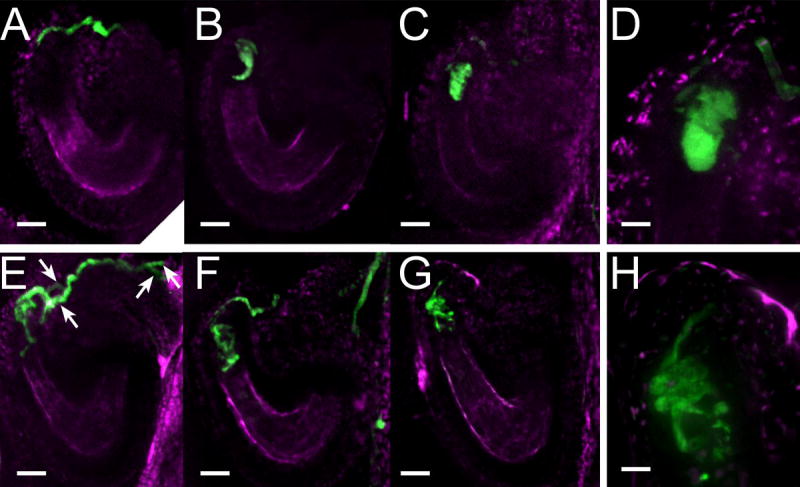
Figure 2. The amc mutation disrupts pollen tube growth arrest and discharge.

(A–H) Examination by confocal microscopy of YFP-expressing pollen 19–24h after manual pollination during wild-type (A–D) and amc mutant (E–H) pollen/ovule interactions. Fluorescence from the YFP expressed in pollen tubes is shown in green while the autofluorescence from the ovule tissues is shown in magenta.
(A) A wild-type pollen tube reaching the micropylar side of the synergid cells.
(B,C) Two examples of wild-type pollen tube discharges. At the extremity of the PT, the fluorescence assumes the shape of the receptive synergid cell in which the pollen tube has released its content.
(D) Close-up of (C).
(E–G) Three examples of continued growth of presumably an amc pollen tube reaching an amc ovule (see Results and Discussion). The mutant pollen tube while reaching the micropylar part of the synergid cells does not discharge. It keeps growing by coiling and branching (F,G) and the pollen tube tips can be spotted past the synergid cells of the mutant ovule (E,F). Among these mutant interactions, two pollen tubes can be frequently observed within a presumed amc ovule (E, white arrows).
(H) Close-up image of the coiled pollen tube shown in (G). Note how the pollen tube coils and branches making the fluorescence outlines rougher than in (D).
Scales bars are 23 μm for A–C, E–G and 7 μm for D,H.
