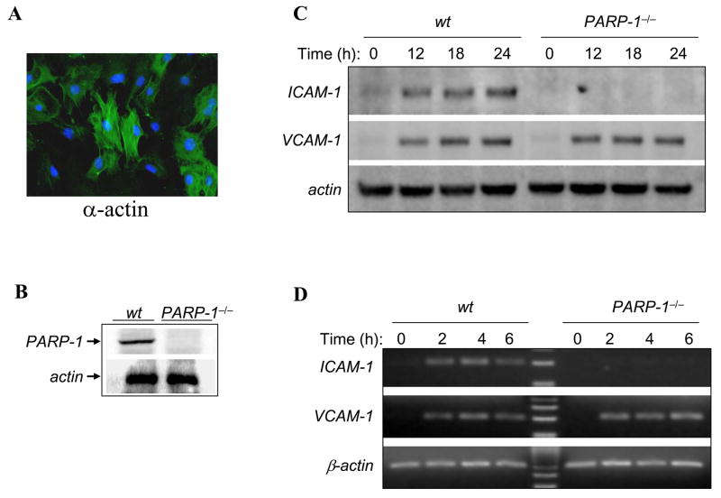
Fig. 1.
Differential expression of ICAM-1 and VCAM-1 in Wt or PARP-1−/− SMCs after TNF exposure. (A) Isolated SMCs were fixed and subjected to immunofluorescence with antibodies to smooth muscle actin (α-actin). (B) SMCs derived from Wt or PARP-1−/− mice were subjected to protein extraction and immunoblot analysis with antibodies to mouse PARP-1 or actin. (C) Wt or PARP-1−/−SMCs were treated with 15 ng/mL TNF for the indicated time intervals after which cell extracts were prepared and subjected to immunoblot analysis with antibodies to mouse ICAM-1, VCAM-1, or actin. (D) SMCs were treated as in (C) but for shorter time intervals after which total RNA was extracted and subjected to RT-PCR using primers specific to mouse ICAM-1, VCAM-1, or β-actin. The amplicons were analyzed by agarose electrophoresis.

