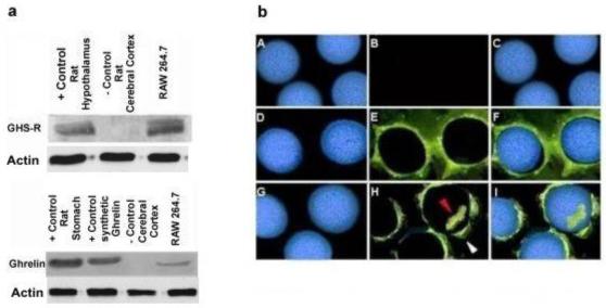
Fig. 1. Ghrelin and GHS-R are expressed constitutively within murine RAW 264.7 macrophages.

Fig 1a; Upper panel demonstrates the presence of GHS-R (Cell lysates from rat hypothalamus/pituitary gland and cerebral cortex were used as positive and negative controls, respectively) and Lower panel shows the presence of ghrelin at protein level (Ghrelin and cell lysates from rat stomach were used as positive controls, while cell lysate from rat cerebral cortex served as a negative controls respectively) in murine macrophage cell line by Western analysis. Fig 1b; Panel (A-C) represents negative controls; Image A is Hoechst staining for nuclei, image B shows no staining in case of negative controls and in image C, latter images A&B have been merged. GHS-R is expressed within RAW 264.7 macrophages as shown by immuno-fluorescence (Panel D-F). Image D is Hoechst staining of the cellular nuclei; image E shows presence of GHS-R on the cell membrane and in image F, latter images D & E have been merged. Ghrelin is co-expressed along with GHS-R on cell membrane and also within cytoplasm (Panel G-I). Image G shows Hoechst staining of nuclei; image H shows the presence of ghrelin on the cell membrane (white arrow) & within cytoplasm (red arrow) and in image I latter images G & H have been merged.
