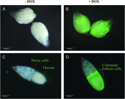
Figure 2.—
DOX-dependent gene overexpression in ovarian follicle cells. Ovaries and stage-10 egg chambers were photographed under a visible light source and fluorescent light source and merged pictures are shown. The scale is indicated in the lower left corner. (A) Ovaries dissected from female containing the GFP-reporter insertion and rtTA(3)E2 insertion cultured in absence of DOX. (B) Ovaries dissected from female containing the GFP reporter and rtTA(3)E2 cultured in the presence of DOX. (C) Stage-10 egg chamber dissected from female containing the GFP-reporter and rtTA(3)E2 cultured in the absence of DOX. (D) Stage-10 egg chamber dissected from female containing the GFP-reporter and rtTA(3)E2 cultured in the presence of DOX. Results are typical of multiple flies and experiments.

