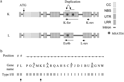
Figure 1.—
Positions of PCR primers and MSATE6 in RGC2 K and RGC2 L. (a) The horizontal lines after K-for and Ex4b indicate the region sequenced and analyzed in this study. (b) Positions of genes TDK and TDL in the RGC2 cluster in cv. Diana are indicated by arrows. Beads in a string indicated the determined physical positions. Genes TDF and TDL were located outside the string. “TD” was omitted for all gene names. Modified from Kuang et al. (2004).

