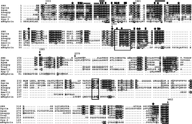
Figure 2.—
Sequence conservation of the SNS cytodomain among various orthologs. Orthologs are shown from the following organisms: Drosophila grimshawi (Dgrim), Anopheles gambiae (Agam), Aedes aegypti (Aaeg), Tribolium castaneum (Tcas), Apis mellifera (Amelli), Caenorhabditis elegans (Syg-2), and mus musculus (mNephrin). Numbered amino acid positions mark the regions of deletion constructs discussed in the text. Stars mark the positions of tyrosine residues and solid squares highlight the serine residues analyzed in this study. Horizontal lines demarcate the putative PDZ-domain-binding sites and boxes surround the putative SH3-domain-binding sites.

