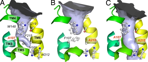
Fig. 4.
Periplasmic pore constriction of AmtB wild type and variants. The solvent-accessible space shown as a light blue surface was calculated by using the program CAVER, with a water-omitted structure. (A) F107A. (B) F215A. (C) F107A/F215A. Selected, highly conserved residues are shown in ball-and-stick representation for the ammonium binding site, phenylalanine gate, and central pore. The substitutions are labeled in red.

