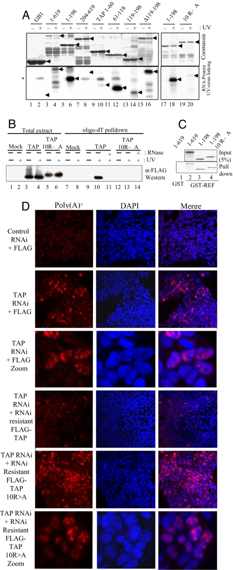
Fig. 4.
Identification of the TAP RNA-binding domain. (A) RNA cross-linking using continuously labeled RNA and GB1-TAP fusions. (B) UV cross-linking mRNP capture assay. 293T cells were transfected with FLAG-TAP-Myc or FLAG-TAP-Myc 10RA vectors and mRNA capture assays carried out after UV irradiation of cells as indicated. TAP proteins cross-linked to the mRNA were detected by Western blotting (WB) with α-Myc Abs. (C) GST pulldowns using GST (lane 1) or GST-REF (lanes 2–4) with GB1-TAP fusions. (D) Rescue of the mRNA export block induced by TAP RNAi. HeLa cells were transfected with pSUPERLUC (control RNAi) or pSUPERTAP (TAP RNAi) together with FLAG, FLAG-TAP, or FLAG-TAP 10RA vectors modified to make the mRNAs resistant to RNAi. Poly(A)+ RNA was detected by using Cy3-oligo(dT)50. The third and sixth sets of panels from the Top show a larger magnification image compared with other panels.

