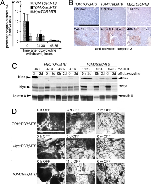
Fig. 2.
Short-term responses to doxycycline withdrawal in mouse mammary tumors induced by Myc and mutant Kras. (A) Decrease in the number of proliferating cells in mammary tumors after doxycycline withdrawal in TOM;TOR;MTB mice (gray bars), TOM;Kras;MTB mice (black bars), and Myc;TOR;MTB mice (hatched bars). Proliferating cells were detected by immunostaining with anti-phosphorylated (Ser-10) histone H3 on paraffin tissue sections and counted as described in Experimental Procedures. (B) Increase in tumor cell death at 24–48 h after doxycycline withdrawal, assayed by immunodetection of anti-activated caspase-3 (Cell Signaling Technology) on paraffin tissue sections from TOM;TOR;MTB mice (Left), TOM;Kras;MTB mice (Center), and Myc;TOR;MTB mice (Right). The slides were counterstained with hematoxylin. Brown-red cells are positive for activated caspase 3. (Upper) Before removal of doxycycline. (Lower) At 24 or 48 h after removal of doxycycline. (Scale bar, 0.5 mm.) (C) Immunoblotting of total lysates from mammary tumors of tritransgenic Myc;TOR;MTB mice (Left) and TOM;Kras;MTB mice (Right) maintained on doxycycline for 7 weeks (0 h OFF) and contralateral tumors harvested 48–50 h after doxycycline removal (2 d OFF). The blot was probed with anti-Myc, anti-Kras, and anti-keratin-8 antibodies as described in Experimental Procedures. (D) Mammary whole mounts from tritransgenic animals taken off the doxycycline diet for the indicated periods of time. (Scale bar, 5 mm.)

