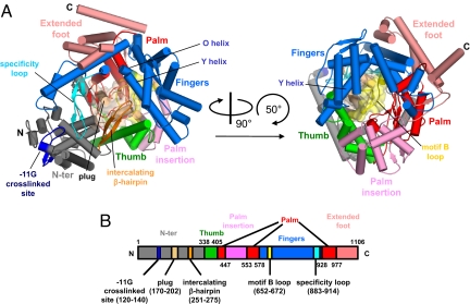
Fig. 1.
X-ray crystal structure of N4 mini-vRNAP. (A) Overall views of N4 mini-vRNAP. Domain and subdomains are represented in their characteristic colors as in B. α-Helices and β-strands are depicted as cylinders and arrows, respectively. The plug and motif B loop are represented as molecular surfaces with partial transparency. Part of the N-terminal domain (residues 120–145, dark blue) has been identified as interacting with the −11 base of the promoter hairpin triloop; this region is a structural counterpart of the AT-rich recognition loop of the T7 RNAP. (Right) Another N4 mini-vRNAP view, derived from (Left) as indicated by the arrows. (B) The thick bar represents the N4 mini-vRNAP primary sequence with amino acid numbering. Domains, subdomains and structural motifs are labeled and color-coded as in A.

