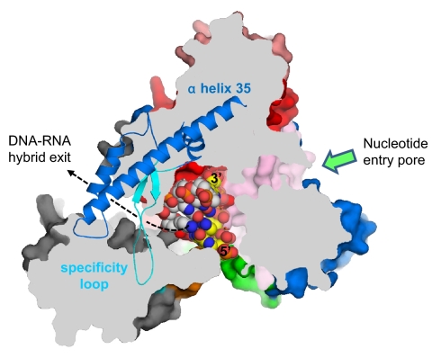
Fig. 2.
A pore for exit of the DNA–RNA hybrid. View of the N4 mini-vRNAP structure, shown as a molecular surface. Orientation of this view is the same as in Fig. 1A Left with the same color code. The molecular surface has been sliced by a parallel plane to this view. The plug module has been removed for clarity. A portion of the Fingers (residues 803–928), which is above the plane, is depicted as a ribbon model, revealing a pore surrounded by the N-terminal domain, Palm core, and Fingers including α-helix 35 and the specificity loop. The pore is ≈25 Å high and ≈15 Å wide. Four base pairs of DNA–RNA hybrid from the T7 RNAP elongation complex (the N4 mini-vRNAP and T7 RNAP elongation complex were aligned at their Palm cores) have been placed in the structure. DNA template strand, white; RNA, yellow; 5′ and 3′ ends of RNA are labeled (5). The RNA extends from the active center to the pore, suggesting that the pore is a putative DNA–RNA hybrid exit channel in the transcription elongation complex; the direction of RNA extending to the pore is indicated by a dashed line. The nucleotide entry pore to the RNAP active center is also indicated by a green arrow.

