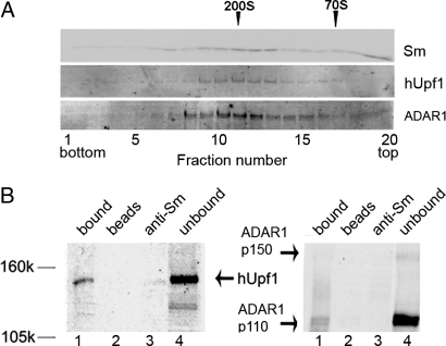
Fig. 1.
hUpf1 is associated with supraspliceosomes. (A) hUpf1 sediments with supraspliceosomes. Supraspliceosomes prepared from HeLa cell nuclei as described in refs. 28 and 29 were fractionated in a sucrose gradient. The 200S fractions were refractionated in a second sucrose gradient. Aliquots from each fraction were run on an SDS/PAGE and were Western blotted with anti-Sm, anti-hUpf1, and anti-ADAR1 antibodies. The sedimentation of 200S TMV and 70S bacterial ribosomes size markers are indicated above the top row. (B) Indirect IP of hUpf1 and ADAR1 from supraspliceosomes. Supraspliceosomes were immunoprecipitated by anti-Sm antibodies, and the precipitated and unbound proteins were analyzed by SDS/PAGE and probed by anti-hUpf1 (Left) and ADAR1 (Right) antibodies (lanes 1 and 4, respectively). As controls, we show the reaction without the antibody (lane 2) and the reaction with antibodies and buffer instead of the sample (lane 3).

