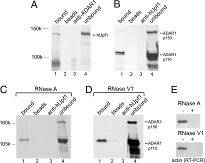
Fig. 3.
ADAR1 and hUpf1 are associated in HeLa cell NE. (A) HeLa cell NE was immunoprecipitated by anti-ADAR1 antibodies, and the precipitated and unbound proteins were analyzed by SDS/PAGE and probed by anti-hUpf1 antibodies (lanes 1 and 4, respectively). Controls: lane 2, no antibody; lane 3, no sample. (B) In a complementary experiment, we show IP of HeLa cell NE by anti-hUpf1 antibodies and Western blotting by anti-ADAR1 antibodies. (C) HeLa cell NE was subjected to RNase A treatment and was then immunoprecipitated by anti-hUpf1 antibodies and Western blotted by ADAR1. The lanes correspond to those in A. (D) The same as in C, except that the NE was treated with RNase V1. (E) RT-PCR of actin RNA extracted from the HeLa cell NE treated and untreated with RNase A (Upper) and RNase V1 (Lower) shown in C and D.

