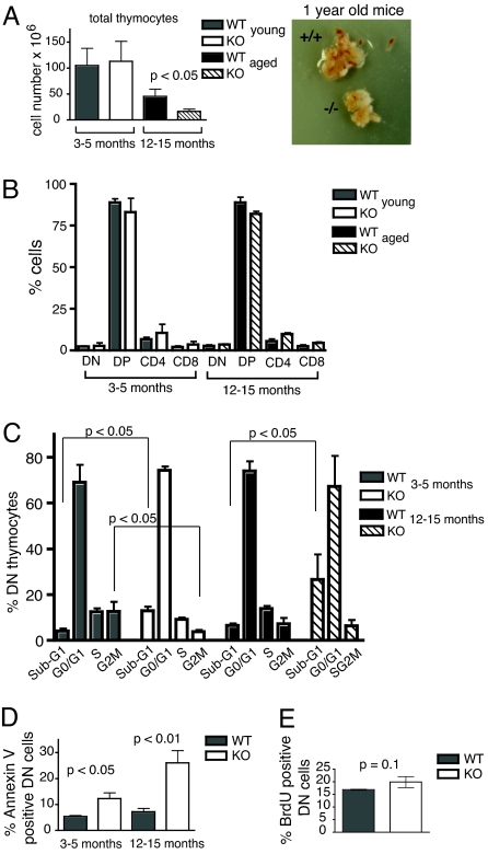
Fig. 1.
Increased DN thymocyte apoptosis and accelerated thymic atrophy in TPPII KO mice. (A) Total thymocyte numbers for young and elderly KO and WT mice (Left) and examples of thymi of 1-year-old littermates (Right). (B) Proportions of major thymocyte subsets determined by staining with anti-CD4 and anti-CD8. In A and B, n = 11 (for young) and n = 6 (for elderly) pairs of littermates, respectively. (C) Cell cycle distribution of DN thymocytes directly ex vivo. DNA staining was performed with propidium iodide. In elderly KO mice, an accurate assessment of S and G2M phase cells could not be made. (D) Annexin V staining of DN thymocytes directly ex vivo. In C and D, n = 3–4 experiments with pooled thymocytes from two or three mice each. (E) In vivo BrdU incorporation into DN thymocytes 1 h after injection of BrdU into 3- to 5-month-old WT or KO mice (n = 3–4 mice per group).

