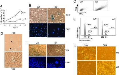
Fig. 4.
Premature senescence of TPPII-deficient fibroblasts and CD8+ T cells. (A) Proliferation of skin fibroblasts derived from newborn mice at passage 10. (B) Microscopic analysis revealed increased numbers of senescent KO cells. Note the flattened, rounded, and enlarged KO fibroblasts with increased expression of β-gal and the larger nuclei as visualized by DAPI staining. (C) Increased cell size of TPPII KO fibroblasts shown by forward scatter/side scatter (FSC/SSC) analysis. (D) Binucleated KO fibroblasts observed by microscopy. (E) Higher percentage of cells with 4N DNA content. (F) Giant multinucleated KO fibroblasts. In all panels, representative experiments including different cell lines at low passage numbers (passage 4–10) are shown. Four different pairs of skin fibroblast lines (three from newborn mice and one from adult mice) were analyzed. (G) Premature senescence of CD8+, but not CD4+, TPPII-deficient splenocytes visualized by staining of acidic β-gal after 4 days of stimulation on anti-CD3. A total of 28 ± 4% of CD8+ KO cells were β-gal+ (n = 3 experiments).

