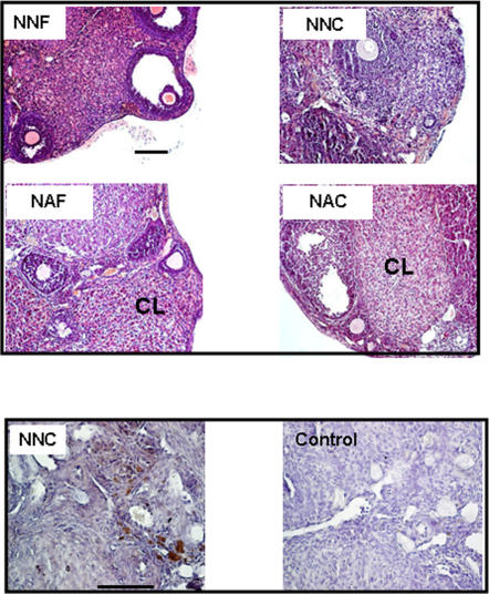
Figure 1. Histological analysis of ovary after one month of grafting and vascularization analysis.
Immature ovarian cortex was grafted in non-pubertal mice freshly (NNF) either after cryopreservation (NNC) or in adult mice freshly (NAF) or after cryopreservation (NAC). Corpus luteum (CL) signs ovulation process. All images are at the same magnification. Representative slide of one ovary from NNC group is shown after immunohistochemical detection of vascularization using α-actin smooth muscle antibody as compared with control (without primary antibody). Scale bar, 100 µm

