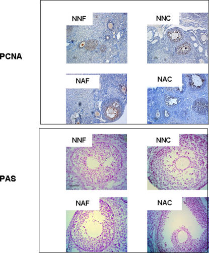
Figure 2. Follicular proliferation and apoptosis.
Cell proliferation was studied using PCNA antibody and apoptosis using PAS staining one month after grafting. Immature ovarian cortex was grafted in non-mature mice freshly (NNF) or after cryopreservation (NNC) or in adult mice freshly (NNF) or after cryopreservation (NNC). All images are at the same magnification. Scale bar, 50 µm

