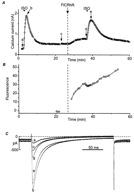
Figure 1. Effect of FlCRhR diffusion on ICa in frog ventricular myocytes.

A, time course of the amplitude of ICa measured every 8 s at 0 mV (holding potential, -80 mV). The myocyte was first exposed to control intracellular and extracellular solutions. A brief (10 s) application of isoprenaline (ISO, 1 μm) induced a large transient increase in ICa. After ICa had returned to basal amplitude, FlCRhR was ejected in the pipette tip at the time indicated by the vertical dashed line, which induced a small rise in ICa. The myocyte was then exposed again briefly (10 s) to ISO. B, average fluorescein and rhodamine fluorescence intensity measured on the cell, indicating diffusion of FlCRhR into the cytosol. C, superimposed ICa current traces obtained at the times indicated by the corresponding letters in A.
