
Figure 1. Propagation of spontaneous increases in [Ca2+]i in the axial direction of the bladder smooth muscle bundles.
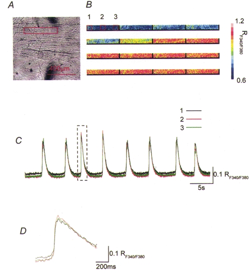
Changes in [Ca2+]i were recorded from an area along the bundle of bladder muscle. A rectangle in the micrograph of the bladder smooth muscle bundle indicates the area where [Ca2+]i recordings were made (A). A series of frames demonstrates that increases in [Ca2+]i concurrently originate along the boundary of the muscle bundle (B). When changes in [Ca2+]i were recorded from three subareas which were located axially by a distance of 100 μm to each other, transient increases in [Ca2+]i occurred in all three areas (C). The onsets and peaks of transient increases in [Ca2+]i occurred almost simultaneously in all three areas (D). A rectangle in C indicates parts of recording traces which are shown in D with a fast time scale.