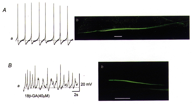
Figure 8. Morphological properties of the communication between smooth muscle cells of the bladder.

The upper membrane potential recording of spontaneous action potentials was obtained using a microelectrode filled with neurobiotin (Aa). The preparation was viewed using a confocal microscope and it was found that neurobiotin readily spread to neighbouring cells located in the axial direction but not to those located in a transverse direction (Ab). The lower membrane potential recording of spontaneous action potentials was obtained from cells which had been exposed to 18β-GA (40 μM) using a microelectrode filled with neurobiotin (Ba). The preparation was again viewed using a confocal microscope and it was found that spread of neurobiotin to neighbouring cells was inhibited by 18β-GA (Bb). The scale bar located to the right of Ba refers to both membrane potential recordings. The calibration bars on Ab andBb represent 100 μm.
