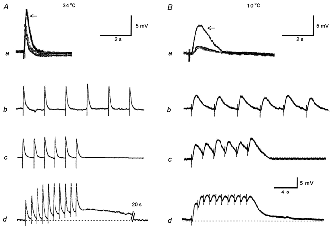
Figure 5. Effect of temperature on excitatory junction potentials (EJPs) and slow depolarizations in hamster tibial artery smooth muscle cells during hibernation.

Aa and Ab, representative traces showing superimposed EJPs to a single stimulus (supramaximal voltage, 0.5 ms) from a control, a cold control and a hibernated animal (arrow) at 34°C (Aa) and 10°C (Ba). The traces from control and cold control animals were nearly identical. Ab-d and Bb-d, membrane responses to electrical field stimulation (supramaximal voltage, 0.5 ms, 6-10 pulses) from hibernated animals at 0.25 Hz (Ab and Bb), 0.5 Hz (Ac and Bc) and 1 Hz (Ad and Bd) at 34°C (A) and 10°C (B). Note that at both temperatures the amplitude of the EJPs from hibernated animals (arrow) are greater than those of control and cold control animals (Aa and Ba), and at 10°C as the frequency progresses (Bd), summations appear which were not present at 34°C (Ad). At 1 Hz at 34°C a slow depolarization emerges (Ad). Each vertical bar shows an electrical stimulus. Resting membrane potentials for Aa and Ba were -62 (control), -61 (cold control) and -60 mV (hibernated) at 34°C, and -59 (control), -60 (cold control) and -59 mV (hibernated) at 10°C, and for Ab, Ac, Ad, Bb, Bc and Bd were -64, -62, -62, -59, -60 and -59 mV, respectively. The lowest scale bar refers to Ab-d and Bb-d.
