
Figure 3. Immunohistochemical localization of ryanodine receptors in Purkinje (A) and ventricular cells(B).
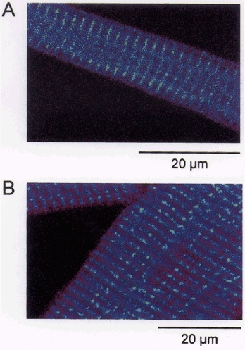
The ventricular cell shows prominent banding at the level of the t-tubules suggesting that ryanodine receptors were located at sites adjacent to the t-tubules (B). The Purkinje cell also exhibited bands of intense staining in the interior of the cell (A). This suggests that ryanodine receptors are located in the cell interior. The spacing between the bands of intensity measured in 4 Purkinje and 4 ventricular cells was found to be 1.95 and 1.98 μm, respectively.