
Figure 3. A working model for the regulation of the facilitated component of glucose absorption by glucose during the assimilation of sugars after a meal.
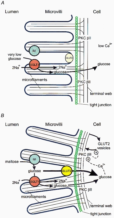
This cartoon of our current working model shows the absorptive epithelial cell before (A) and after (B) a meal. Before a meal, the glucose concentration in the lumen is very low, being less than that in the blood. The level and intrinsic activity of GLUT2 are low, indicated by the pale yellow ellipse for GLUT2; absorption of glucose against its concentration gradient occurs through SGLT1. After a meal, high local concentrations of glucose are present at the microvilli from the hydrolysis of dissacharides, for example, by the action of isomaltase (IM) on maltose. Transport of glucose through SGLT1 results in activation of PKC βII and activation and recruitment of GLUT2 to the brush-border membrane, indicated by the bright yellow circle for GLUT2. The rate of absorption through GLUT2 is then severalfold greater than that through SGLT1. Transport of glucose through SGLT1 also results in contraction of the peri-junctional actomyosin ring (just below the tight junction), causing subtle rounding of the surface of the absorptive cell. Both processes may be mediated by a glucose-induced increase in intracellular Ca2+ concentrations. For further explanation see text.