
Figure 2. Whole-cell currents evoked by kainate and ATPA.
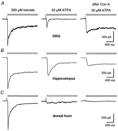
Currents recorded in a freshly dissociated DRG cell (A), a cultured hippocampal neuron (B), and cultured dorsal horn neurons (C). All three cell types were from 1-5 day postnatal animals. Although kainate evoked very similar currents in the 3 cells, responses to ATPA were significantly different in these cells, which are representative of the neurons from each region. The right hand column shows currents evoked by ATPA after exposure to Con A. In A and B, the traces before and after Con A are from the same cell. In C, the two traces before Con A are from a different neuron than the trace after Con A. Exposure to Con A blocked desensitization to kainate in this spinal neuron (not shown), but did not enable ATPA to elicit significant current. Holding potential, -80 mV.