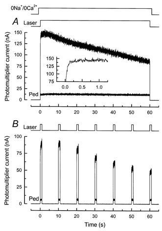
Figure 2. Fluo 5F fluorescence recorded from a salamander rod in 0 Ca2+, 0 Na+ solution.

Fluorescence magnitude is represented by the current recorded from the photomultiplier. A, dark-adapted rod was stepped to 0 Ca2+, 0 Na+ solution (see Methods) 1 s before beginning of laser exposure. Laser shutter was opened for 60 s (laser intensity, 2.5 × 10⁹ photons μm−2 s−1), giving the larger of the two traces. One second after laser exposure, the rod was rapidly returned to Ringer solution; approximately 45 s later, the outer segment was stepped a second time to 0 Ca2+, 0 Na+solution and the laser shutter opened for 60 s to give smaller trace (Ped). Inset, larger of two traces at higher temporal resolution, showing time-dependent increase in fluorescence (compare to inset of Fig. 1A). B, same protocol and laser intensity as in A for another rod, but instead of continuous laser illumination the rod was repeatedly exposed to the laser for 1 s at 10 s intervals, effectively reducing the total exposure of the rod to the laser by a factor of 10. Larger signals were recorded from dark-adapted rod; smaller signals (Ped) from a second presentation after return of the outer segment to Ringer solution.
