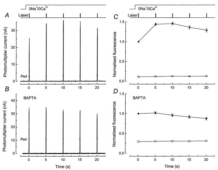
Figure 5. Effect of BAPTA incorporation on fluo-5F responses to brief laser flashes in rods exposed to 0 Ca2+, 0 Na+ solution.

Laser intensity, 2.4 × 10¹⁰ photons μm−2 s−1. A and B, show sample fluorescence responses from dark-adapted rods in 0 Ca2+, 0 Na+ solution in the absence and presence of BAPTA. C and D show mean ±s.e.m. under equivalent conditions from rods dissociated from the same animal. A, protocol identical to Fig. 3A: 5 laser flashes of 100 ms duration were delivered at 5 s intervals commencing 1 s after stepping to 0 Ca2+, 0 Na+ solution. Pedestal values measured 45 s after return to Ringer solution are indicated by the bars (Ped). B, protocol as for A but fluo-5F incubation solution included 50 μm BAPTA AM. Pedestal values are indicated by the bars (Ped). C, mean ±s.e.m. for 6 rods as in A (without BAPTA). For each cell the fluorescence signal has been normalised to the fluorescence elicited by the first laser flash. First exposure of dark-adapted rod to laser flashes in 0 Ca2+, 0 Na+ solution (•); pedestal values (○), for which the standard errors were smaller than the symbol width. D, mean ±s.e.m. for 8 rods as in B (after BAPTA incorporation). Note much smaller fluorescence increase and somewhat slower decline. Larger relative magnitude of pedestal values suggests either that BAPTA buffering has altered rod [Ca2+]i in darkness or that light-induced Ca2+ release is underestimated with this pulse protocol, or both.
