
Figure 6. The effect of 5-HT on presynaptic action potentials.
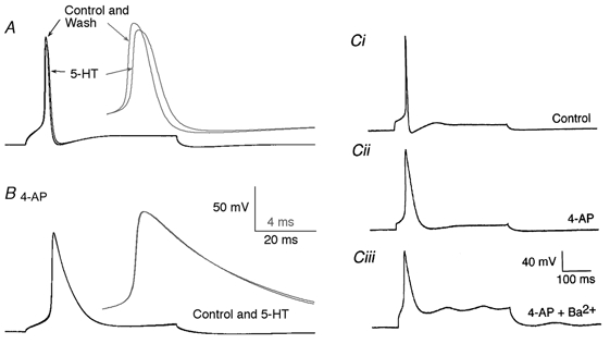
Action potentials in the reticulospinal axons were recorded with microelectrodes under current clamp. A, an action potential evoked by a depolarizing current pulse (0.5 nA). Application of 5-HT (30 μm) reduced the amplitude of the evoked action potential. The inset (grey) shows the traces on an expanded time scale. (Note that the peak of the action potential was reduced in amplitude.) B, an action potential was recorded in the same axon in the presence of 4-AP (30 μm). Application of 5-HT had no effect. The inset (grey) shows the traces on an expanded time scale as in A. Ci, another example of an action potential recorded under control conditions. An action potential was evoked in a different axon by a 1 nA depolarizing current pulse. Cii, the same axon as in Ci after the application of 4-AP (30 μm). Ciii, the same axon as in Ci after the replacement of extracellular Ca2+ with Ba2+ and in the presence of 4-AP.