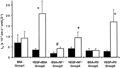
Figure 2. Chronic effect of acute inhibition of Ca2+ influx or MEK on VEGF-mediated increased Lp.

Permeability was measured on day 1 (▪) during perfusion with 1 % BSA alone or 1 % BSA and inhibitor (5 mm Ni2+ or 30 μm PD98059). Vessels were then perfused with BSA (group 1, n = 6), 1 nm VEGF and BSA (group 2, n = 28), VEGF, BSA and the inhibitor (group 4, n = 6 and group 6, n = 8), or with just BSA and the inhibitor (group 3, n = 5 and group 5, n = 5). The following day the Lp was measured on the same vessel (□) during perfusion with 1 % BSA alone. *P < 0.01 compared to day 1, #P = 0.03 compared to day 1, + P < 0.01 compared to vessels exposed to VEGF in the absence of nickel (unpaired t test).
