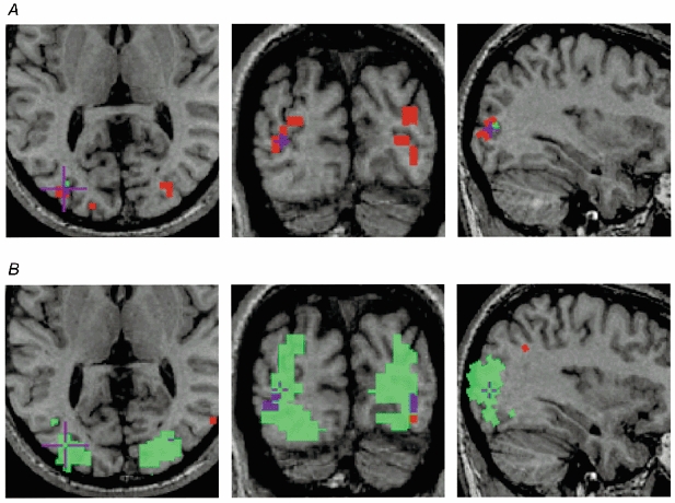
Figure 4. Axial (left), coronal (middle) and sagittal (right) anatomical images of subject JF.

Significant (P < 0.05) functional voxels during fixing (green) and amblyopic (red) eye stimulation are superimposed. Purple is used to show the area of overlap. A, response to the 11 c.p.d. grating. Note that this grating was beyond JF's amblopic eye acuity, yet there was significant activation when this eye was stimulated. B, the response to the 4 c.p.d. grating shows a large area of cortex driven by the fixing eye, yet relatively little detectable activation when the amblyopic eye was stimulated.
