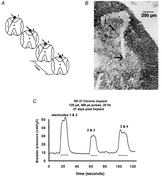
Figure 2. Scheme for chronic microstimulation in the feline sacral spinal cord.

A, the arrays of 4 individual iridium microelectrodes were implanted at rostro-caudal intervals of approximately 1 mm, and directed towards the intermediolateral cell column and the preganglionic parasympathetic nucleus. B, site of the tip of one of the microelectrodes (arrow), close to the PPN (1 μm section of epoxy-embedded spinal cord, stained with Toluidine Blue and Azure II). C, hydrostatic pressure within the urinary bladder induced by stimulating with various pairs of microelectrodes. The stimulus pulses were applied to the 2 electrodes in an interleaved manner. (Modified from Woodford et al. 1996, with permission.)
