
Figure 1. Electrophoresis of MHC isoforms.
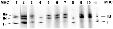
Cell cultures were grown for 28 days without any treatment (lane 9) or from day 14 of the culture onwards for a further 14 days in the presence of cyclosporin A (500 ng ml−1) (lane 10) or the Ca2+ ionophore A23187 (4 × 10⁻⁷m) (lane 3) or cyclosporin A and Ca2+ ionophore (lane 5). Other cultures were electrostimulated (1 Hz for 15 min, stimulus duration 2.5 ms, followed by a pause of 30 min) (lane 2) or electrostimulated and treated with cyclosporin A (lanes 6 and 7) from day 14 onwards for a further 14 days. Myosin extracts of the homogenized cells were separated on SDS polyacrylamide gels (SDS-PAGE). Markers were run in lane 1 (mixture of myosin extracts from m. psoas and m. vastus intermedius, red portion, of the rabbit), and lanes 4, 8, and 11 (mixture of myosin extracts from m. psoas and m. soleus of the rabbit).