
Figure 2. Effect of cyclosporin A, Ca2+ ionophore, and electrostimulation on the expression of slow MHCI mRNA.
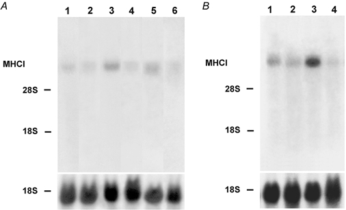
A, cell cultures were grown for 28 days without any treatment (lane 1) or from day 14 of the culture onwards for a further 14 days in the presence of cyclosporin A (500 ng ml−1) (lane 2) or the Ca2+ ionophore A23187 (4 × 10⁻⁷m) (lane 3) or cyclosporin A and Ca2+ ionophore (lane 4). Other cultures were electrostimulated (1 Hz for 15 min, stimulus duration 2.5 ms, followed by a pause of 30 min) (lane 5) or electrostimulated and treated with cyclosporin A (lane 6) from day 14 onwards for a further 14 days. B, cell cultures were grown for 16 days without any treatment (lane 1) or from day 14 of the culture onwards or a further 2 days in the presence of cyclosporin A (500 ng ml−1) (lane 2) or Ca2+ ionophore (4 × 10⁻⁷m) (lane 3) or cyclosporin A and Ca2+ ionophore (lane 4). Total RNA (20 mg) was isolated from control and treated cultures at the time points indicated, fractionated on a 1.2 % formaldehyde agarose gel, and transferred to nitrocellulose. The blots were hybridized with the 32P-labelled 3′ terminal Hin fI fragment of MHCI cDNA or an 18S rDNA probe. The positions of 18S rRNA (1.9 kb) and 28S rRNA (4.8 kb) on the ethidium bromide-stained gel are indicated.