
Figure 6. L-type Ca2+ currents and Ca2+ transients evoked by short control and long post-MI action potentials.
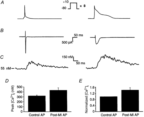
A, the voltage protocols: control (left) and post-MI (right) APs were applied to Cs+-loaded rat ventricular myocytes following a loading train of eight 100 ms steps to +10 mV. B, Ca2+ currents evoked by the control and post-MI APs were measured as Cd2+-sensitive currents and differed in peak amplitude, time to peak and integrated current. Table 1 summarizes the characteristics of ICa,L during control and post-MI APs. C, representative intracellular Ca2+ transients triggered by control and post-MI APs. D, peak systolic Ca2+ was not significantly different between control (319.2 ± 16.1 nm, n = 5) and post-MI APs (433.4 ± 48.2 nm, n = 5). E, normalized peak Ca2+ transients of the post-MI AP were only 1.4-fold greater than the control AP under these intracellular conditions.