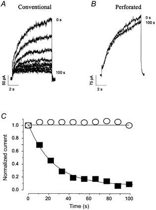
Figure 1. IKs in uninfected neonatal murine ventricular myocytes.

A and B, representative raw current traces of slowly activating time-dependent outward currents (IKs) elicited by long depolarization (8 s) of neonatal mouse myocytes to +70 mV from a holding potential of -40 mV using conventional patch-clamp (A) and perforated-patch (B) techniques. C, current magnitude, measured as the difference in outward current at the beginning and end of the pulse, is plotted against time during these experiments. ▪, conventional; ○, perforated-patch recording. Rapid current run-down was observed when IKs was recorded using the conventional patch-clamp technique but was absent when the perforated-patch technique was employed. Cell capacitance was 42 and 25 pF, respectively, for these cells.
