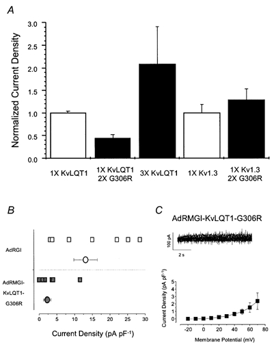
Figure 3. Effects of constructs carrying the dominant-negative mutation G306R.

A, bar graphs summarizing the specific inhibitory effects of transfection of pAdRMGI-KvLQT1-G306 on KvLQT1 channels. For each group of channels, current amplitudes were normalized by the mean current of the same channel type (transfected with 1 × DNA) expressed alone. Current through KvLQT1 channels increased when more pAdRMGI-KvLQT1 DNA was used for transfection but was substantially suppressed when it was co-expressed with pAdRMGI-KvLQT1-G306R. In contrast, co-expression of Kv1.3 channels with KvLQT1-G306R did not lead to current suppression compared to expression of Kv1.3 alone. Currents were measured at the end of a 4 s, +70 mV or 1 s, +60 mV pulse from a holding potential of -40 or -100 mV for KvLQT1 and Kv1.3 channels, respectively. B, distribution of raw (squares) and averaged (circles) current densities at +70 mV recorded from AdRGI- (open symbols) and AdRMGI-KvLQT1-G306R-infected (filled symbols) neonatal mouse myocytes. The latter was significantly (P < 0.05) reduced. C, top, typical current traces of IKs recorded during a family of stepping voltages from AdRMGI-KvLQT1-G306R-infected myocytes. Bottom, I-V relationship.
