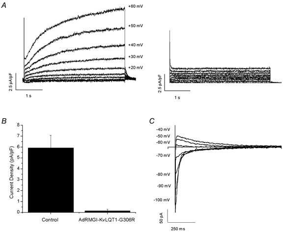
Figure 4. IKs in isolated adult guinea-pig ventricular myocytes.

A, representative raw current traces of IKs recorded from control uninfected (left) and AdRMGI-KvLQT1-G306R-infected (right) myocytes. B, bar graph summarizing the current densities measured at the end of a 4 s depolarizing pulse to +60 mV. C, typical tail currents of IKs recorded from a control uninfected myocyte during a family of stepping voltages after a 4 s prepulse to +70 mV.
