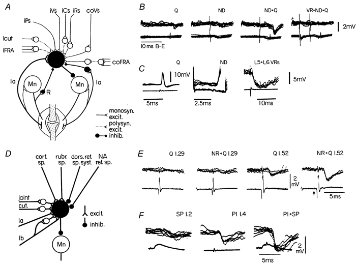
Figure 1. Convergence on interneurones in the pathway of reciprocal Ia inhibition (A-C) and on interneurones in the Ib inhibitory pathway (D -F).

Circuit diagrams in A and D summarise several of the connections to these two types of interneurones. Recordings from a knee flexor motoneurone (B) demonstrate the spatial facilitation on combined stimulation of quadriceps (Q) Ia afferents and Deiters' nucleus (ND). The right-most records show the reduction of the IPSP when conditioned by the L5 + L6 ventral roots (recurrent inhibition, cf. circuit diagram in A). Direct recording from a Ia inhibitory interneurone (C) demonstrates the monosynaptic excitation from Q and ND and the disynaptic recurrent inhibition following stimulation of the L5 + L6 ventral roots. Recordings in E demonstrate the facilitatory interaction between a conditioning rubrospinal volley (NR) and a group I volley from Q. Note that the facilitation is only seen when the quadriceps volley is strong enough to activate Ib afferents (stimulation strength of 1.52 × threshold (T) for recruiting the most excitable nerve fibres). Recordings in F demonstrate the facilitatory interaction between cutaneous fibres (superficial peroneal nerve, SP) and group I afferents from the nerve to the plantaris muscle (Pl). Upper traces are intracellular records (voltage calibrations apply to these traces), while lower traces are incoming volleys recorded from the dorsal root entry zone. (B adapted from Hultborn & Udo, 1972; C from Hultborn et al. 1976; E from Hongo et al. 1969; F from Lundberg et al. 1977.)
