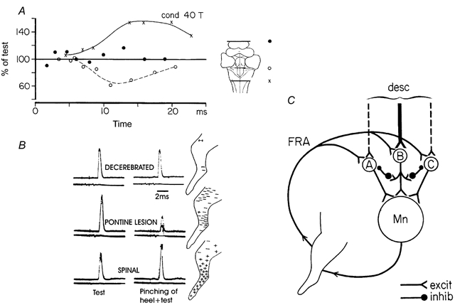
Figure 2. Differential release of transmission in excitatory and inhibitory ‘flexor reflex afferents’ by brainstem lesions and spinal transection.

The graph in A shows the time course of the effects of single conditioning volleys in the nerve to flexor digitorum longus (40 T) on monosynaptic reflexes evoked from knee flexor posterior-biceps and semitendinousus. In B the effects of pinching the heel was tested under similar conditions. C, diagram showing alternative reflex pathways from flexor reflex afferents (FRA) with descending excitatory connections to interneurones of these pathways and its inhibitory interactive connections with the other reflex pathways from FRA. (A and B adapted from Holmqvist & Lundberg, 1961; C adapted from Lundberg, 1973.)
