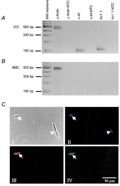
Figure 1. Transcriptional expression of Kv1.1 in isolated smooth muscle cells of murine fundus and isolated murine fundus IC-IM.

Phase and fluorescence photomicrographs of Kv1.1-LI and c-Kit-LI in isolated murine fundus cells. RNA was prepared from isolated ICC. A, freshly dispersed murine fundus IC-IM. B, freshly dispersed murine fundus myocytes. c-kit and Kv1.1 were expressed in ICC but not in SMCs. Kv1.1 expression in ICC was confirmed at the protein level. C, phase contrast and fluorescence photomicrographs of a freshly dispersed ICC and a smooth muscle cell isolated from murine fundus. Panel I, a phase contrast photomicrograph of an ICC (arrow) and a smooth muscle cell (arrowhead). Panel II, photomicrograph of Hoechst 33342 fluorescence (blue) showing viable nuclei in both the ICC (arrow) and the smooth muscle cell (arrowhead). Panel III, photomicrograph of c-Kit-LI fluoresecence (red) in the ICC (arrow) but not in the smooth muscle cell. Panel IV, photomicrograph of Kv1.1-LI fluorescence (green) in the ICC (arrow) but not in the smooth muscle cell.
