
Figure 9. Two classes of back propagation.
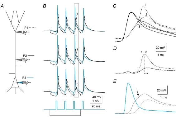
A, schematic diagram showing the placement of 3 electrodes on a L5 pyramidal neuron simultaneously. B, a train of back-propagating APs was elicited by current injections at the soma at 70 Hz with progressively larger hyperpolarizing current (-600 to -700 pA) injected at the proximal electrode (300 μm from the soma). (Somatic recording, P3, blue; proximal recording (300 μm), P2, black; and distal recording, 630 μm, P1, dashed line.) The hyperpolarization eventually caused the third AP in the train to decrease in amplitude, revealing the presence of a mechanism maintaining the amplitude of the back propagation of APs. C, expanding the time scale for the boxed region in B and overlaying the traces shows the two-component nature of the back-propagating AP at the distal site just before inactivation (1), during (2) and after full inactivation (3), black traces. A small depolarization is visible at the proximal site (black traces) following the peak at the proximal site. D, by subtracting trace 3 from trace 1, the regenerative component of the back-propagating AP is revealed to arise first at the distal location and reflected back towards the soma. Peaks are shown by vertical lines. E, a different cell in which the back-propagating AP clearly had two components under control conditions. Recordings were made at 170 μm (dashed trace), 420 μm (black trace) and 630 μm (blue trace) from the soma. The point of inflection revealing the existence of a further regenerative component on the recording from the 420 μm site is shown by an arrow.