
Figure 3. Inwardly rectifying K+ currents in 60 mm external K+.
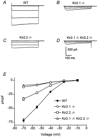
A–D, inwardly rectifying currents were recorded in 60 mm external K+ from voltage-clamped neonatal myocytes isolated from wild-type, Kir2.1−/−, Kir2.2−/− and Kir2.1−/−-Kir.2.2−/− mice. For each family of current traces, the voltage was stepped from a holding potential of −10 mV to potentials of −10 to −70 mV (in 15 mV increments). Leak currents were subtracted using currents evoked by small depolarizing pulses (P/4 method). Cell capacitances were 15, 14, 11 and 15 pF, respectively. E, steady state currents (means ±s.e.m.) were plotted as a function of voltage in wild-type (n = 4), Kir2.1−/− (n = 8), Kir2.2−/− (n = 11) and Kir2.1−/−-Kir.2.2−/− (n = 9) myocytes. Current amplitudes were measured at the end of the 500 ms voltage-clamp step and were normalized to cell capacitance to control for cell surface area.