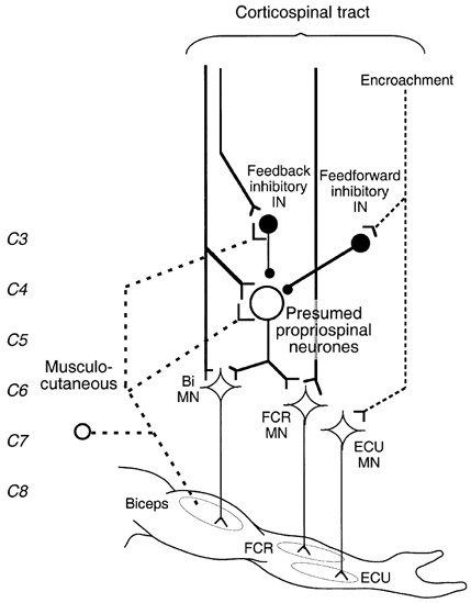
Figure 9. Wiring diagram of possible connections investigated in the present studies.

The thick dashed line shows the pathway mediating non-monosynaptic group I excitation from musculo-cutaneous to biceps (Bi) and flexor carpi radialis (FCR) motoneurones (MNs). Y-shaped bars and small filled circles represent excitatory and inhibitory synapses, respectively. The large open circle represents excitatory propriospinal neurones (PNs) with diverging projections onto Bi and FCR MNs. Medium sized filled circles represent inhibitory interneurones: feedback inhibitory interneurones fed by both musculo-cutaneous afferents and corticospinal projections, and feedforward inhibitory interneurones fed only by corticospinal projections. The corticospinal tract (thick vertical lines) is shown activating Bi and FCR MNs through a monosynaptic projection and a disynaptic connection through the presumed PNs (which can be inhibited by feedback and feedforward inhibitory interneurones), and separately activating feedback inhibitory interneurones. High TMS intensities lead to widespread activation of ‘inappropriate’ (encroachment) corticospinal projections (vertical dashed line) activating, e.g. extensor carpi ulnaris (ECU) MNs and feedforward inhibitory interneurones directed to PNs fed by biceps afferents.
