
Figure 1. Properties of GABA currents recorded in somata and dendrites.
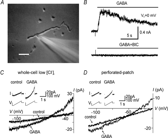
A, photograph of a dendritic recording. The recording electrode is shown on the right. Scale bar, 20 μm. B, currents recorded in a dendrite in perforated-patch mode in response to local superfusion with 2 μm GABA (top) and GABA in combination with 40 μm bicuculline (BIC; bottom). The holding potential (Vh) was 0 mV. The horizontal bars indicate the duration of the drug applications. C, ramp currents and corresponding I-V relationships recorded in conventional whole-cell mode in a dendrite in control solution or 2 μm GABA. Currents were recorded in response to a voltage ramp from −110 to −30 mV from a holding potential of −60 mV (inset). This record was obtained with a low Cl− (9 mm), Cs+-based patch solution. D, ramp currents and corresponding I–V relationships recorded in a different dendrite in gramicidin perforated-patch mode. Note the similar shape and outward rectification of GABA currents in C and D.