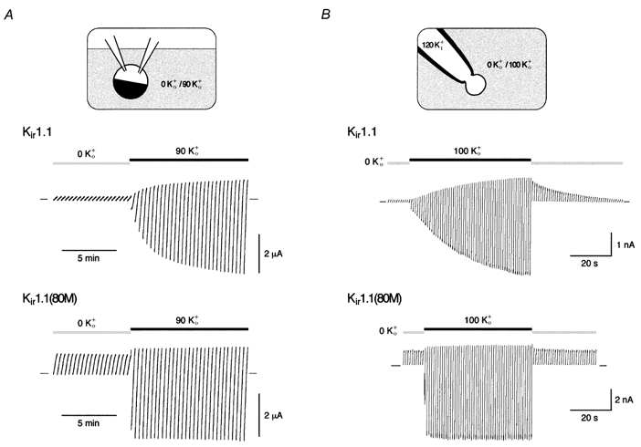
Figure 1. K+-dependent gating of Kir1.1 channels.

A, currents mediated by Kir1.1 (upper trace) or Kir1.1(K80M) channels (lower trace) recorded in oocytes preincubated in K+-free solution for more than 2 h. Change in bath solution from 0 K+o to 90 K+o as indicated by horizontal bars; currents were recorded in response to voltage ramps from −120 to 50 mV (in 20 s), and zero current is indicated by small bars. B, currents recorded in giant outside-out patches from oocytes expressing Kir1.1 (upper trace) or Kir1.1(K80M) channels (lower trace) and treated as in A. Currents are response to voltage ramps from −100 to 50 mV (in 400 ms) at pHi 7.1; application of extracellular solutions as indicated. Note the difference between slow increase in Kir1.1 currents due to recovery from K+-dependent inactivation and rapid increase in current amplitude due to the shift in EK resulting from the change in [K+]o.
