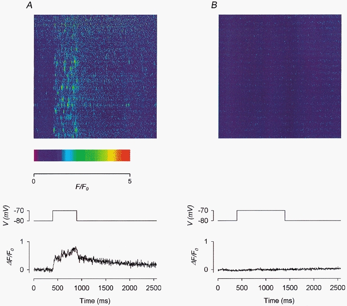
Figure 8. Effect of 20 μm Ry on depolarization-evoked calcium sparks and global calcium transients.

A, control before the application of Ry. B, in the presence of 20 μm Ry. Each panel shows: top, a line-scan image; middle, the depolarizing pulse used to trigger the calcium release; bottom, the global calcium transient trace obtained by compressing the 2-D image vertically into a 1-D array. Fibre 14. Holding potential −80 mV. Sarcomere length 3.7 μm.
